
The carotid artery is one of the most important arteries in the human body. In fact, there are two carotid arteries, one on each side of the neck, which are responsible for carrying oxygen-rich blood from the heart to the brain. Smooth blood flow through each carotid artery is vital, as the brain is completely dependent on this supply to function properly.
When a carotid artery becomes narrowed, it means that the inner wall of the vessel has become narrowed due to the buildup of plaque. Plaque is made up of cholesterol, calcium, cellular debris, and fibrin. This process, known as atherosclerosis, restricts blood flow. Even more dangerous is the possibility that a small piece of this plaque, or a clot, could break off and travel to the brain, causing an ischemic stroke. Understanding when this condition requires surgical or endovascular intervention is key to preventing permanent damage.
Symptoms and risk of stroke
Carotid stenosis is usually a "silent" condition. This means that the patient does not feel pain or have any obvious discomfort, so the diagnosis is often made incidentally during a preventive check-up (such as a triplex cervical ultrasound).
However, when the narrowing in the carotid artery is advanced or a portion of the plaque becomes unstable, symptoms may occur. These are directly related to a transient ischemic attack or a full-blown stroke. The most common symptoms include:
- Sudden weakness or numbness in the face, arm, or leg, usually only on one side of the body.
- Difficulty speaking (dysarthria) or sudden inability to understand what others are saying.
- Sudden loss of vision or blurred vision in one eye (a condition called transient amaurosis).
- Loss of balance, dizziness, or sudden difficulty walking without an obvious cause.
Every second counts when these symptoms appear, as early medical evaluation can prevent permanent disability.
How is the degree of stenosis diagnosed and measured?
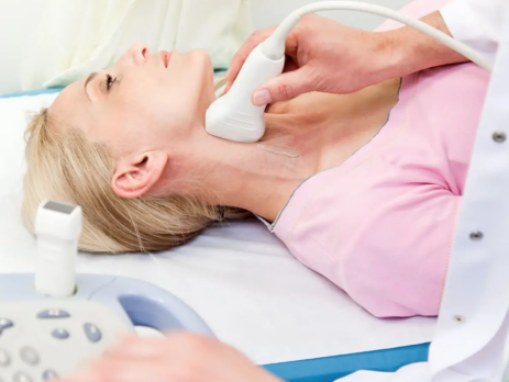
Accurate diagnosis is the first and most critical step in determining the appropriate treatment strategy. The main test of choice for evaluating the condition is a color ultrasound (Triplex) of the carotid arteries. This is a painless, non-invasive test that allows the doctor to see the structure of the vessel and measure the speed of blood flow. This speed increases when blood is forced to pass through a narrower point, allowing the calculation of the degree of stenosis.
If Triplex shows significant damage, the vascular surgeon usually orders further imaging to confirm the findings and plan the procedure. These tests include CT angiography or magnetic resonance angiography (MRA). In some more complex cases, digital subtraction angiography may be required, which offers the most detailed visualization of the vascular anatomy, although it is a more invasive method.
When does carotid stenosis require surgery?
The decision on whether a patient should undergo surgery is based on guidelines from international medical societies, such as the European Society of Vascular Surgery and the American College of Cardiology. Patients are divided into two broad categories: symptomatic and asymptomatic.
For symptomatic patients, that is, those who have had a stroke or transient ischemic attack within the past six months, surgery is almost always recommended when the carotid stenosis exceeds 70%. In many cases, surgery is even considered for stenosis between 50% and 69%, if the atherosclerotic plaque is considered particularly unstable or progressive. Surgery in these patients should ideally be performed within the first two weeks of the episode to maximize the benefit of preventing a new, more severe stroke.
For asymptomatic patients, the criteria are stricter. Surgery is usually considered when carotid stenosis reaches or exceeds 70% to 80%, and only if the patient is at high risk of stroke despite optimal medical treatment, or if their life expectancy allows them to benefit from the procedure in the long term.
The available surgical and endovascular methods
When it is deemed necessary to restore the patency of the carotid artery, there are two main therapeutic approaches. The choice depends on the anatomy of the vessel, the patient's age, their general health status, and the experience of the surgical team.
The first and most proven method is carotid endarterectomy. This is an open surgical procedure performed under general or local/regional anesthesia. The surgeon makes a small incision in the neck, gains access to the carotid artery, carefully opens the vessel, and removes the atherosclerotic plaque that is blocking the flow. The vessel is then closed, often with the use of a special stent to prevent future restenosis.
Dr. med. Nektarios Galanis ' team performs this procedure under local anesthesia. This allows them to monitor the patient's brain function throughout the surgery and intervene if necessary to prevent an intraoperative stroke. Dr. med. Nektarios Galanis is one of the few Vascular Surgeons who perform this procedure.
The second method is endovascular repair with stent placement (carotid stenting). Although this technique is less invasive, it has a higher rate of intraoperative strokes than open surgery and is preferred in special cases, such as people who have previously undergone radiation to the neck. The specialist doctor inserts a thin catheter, usually from the thigh (femoral) or arm (radial) artery, and guides it to the area of stenosis. There, a special metal mesh (stent) is deployed that presses the plaque against the walls and keeps the vessel open, ensuring the free passage of blood.
Conservative treatment and lifestyle changes
Even when carotid stenosis does not meet the criteria for surgery, medical monitoring and conservative treatment remain mandatory. The goal of conservative treatment is to stabilize the atherosclerotic plaque and prevent further narrowing of the vessel lumen.
Medication usually includes antiplatelet drugs (such as aspirin or clopidogrel) that prevent clot formation, as well as statins. Statins not only reduce blood cholesterol levels, but also have a very important pleiotropic effect: they stabilize the plaque wall, making it less prone to rupture and detachment.
At the same time, lifestyle changes play a decisive role in the progression of the disease. Smoking cessation is the most immediate and effective intervention, as smoking dramatically accelerates atherosclerosis. Regulating blood pressure and diabetes, adopting a balanced Mediterranean diet with low fat and regular aerobic exercise help maintain the health of the vascular system and protect the brain from future risks.